Best Hospital in Ghatkopar, Mumbai | Zynova Shalby Hospital


Blogs Zynova
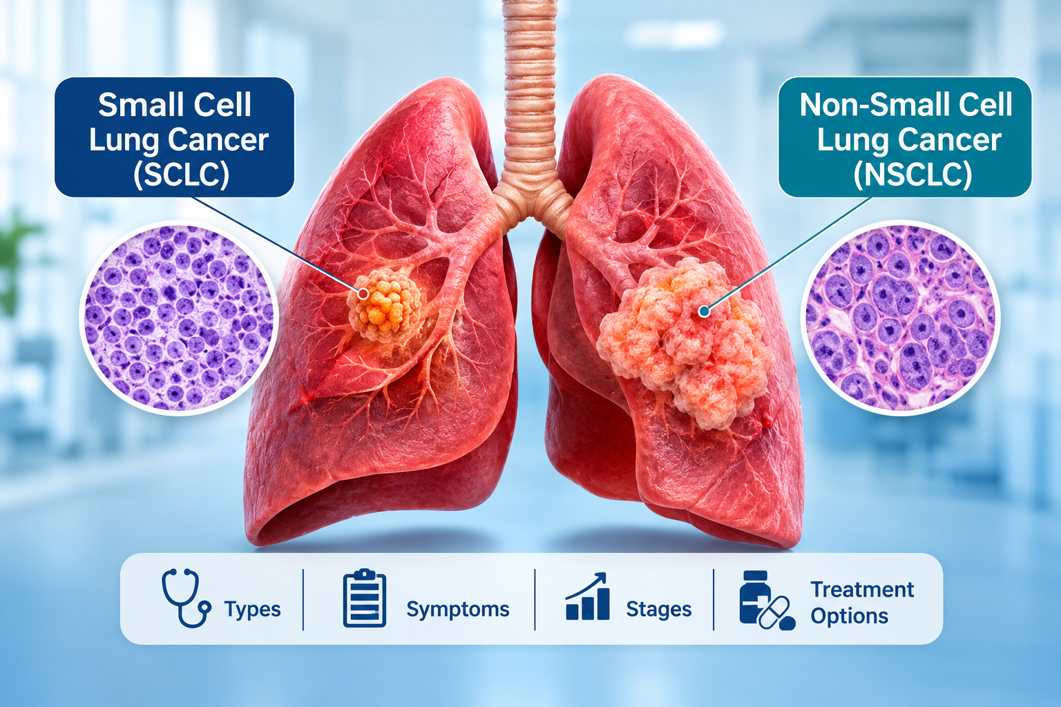
Small Cell vs Non-Small Cell Lung Cancer: Types, Differences and Treatment Options
When a doctor says “lung cancer”, the next question is almost always “which type?” Everything after that depends on the answer: the tests you need, the treatment you are offered, and how fast decisions have to be made. There are two main kinds. Small cell lung cancer and non small cell lung cancer look different under a microscope, grow at different speeds and respond to different treatments. This guide explains small cell vs non small cell lung cancer in plain language, covering symptoms, stages, diagnosis and treatment. If you want the wider picture first, our article on lung cancer in India is a good place to start. What Is Lung Cancer? Lung cancer begins when cells in the lung change and start multiplying without the usual controls. They can form a lump, press on nearby airways and blood vessels, and sometimes spread through the lymph nodes or bloodstream to other organs. Doctors sort the types of lung cancer by what the cells look like. That gives two groups: small cell lung cancer (SCLC), about 10 to 15 percent of cases, and non-small cell lung cancer (NSCLC), about 80 to 85 percent. Knowing the group is not a formality. It decides whether surgery is realistic, whether a tablet-based targeted treatment could work, and whether treatment needs to begin within days. What Is Small Cell Lung Cancer (SCLC)? Small cell lung cancer takes its name from the small, round cells a pathologist sees in the sample. You may also hear it called oat cell carcinoma. It usually starts in the large airways near the centre of the chest. How fast does SCLC grow and spread? Fast. SCLC cells divide quickly and often travel early to lymph nodes, the liver, bones, adrenal glands and brain. Many people already have spread beyond the chest by the time it is found. The upside of that fast growth is that SCLC often responds well to chemotherapy and radiation at first. The difficulty is that it can come back, so follow-up matters. Key features of SCLC It is very strongly linked to tobacco and is uncommon in people who have never smoked. It is a neuroendocrine cancer, so it can sometimes upset hormone and salt levels. A rarer form, combined small cell lung cancer, contains both small cell and non-small cell areas. Surgery is used only in a small number of very early cases. What Is Non-Small Cell Lung Cancer (NSCLC)? Non small cell lung cancer is a group, not a single disease. It covers every lung cancer that is not small cell. As a rule it grows more slowly than SCLC, and more cases are found while still inside the chest, which keeps more treatment options open. Adenocarcinoma The most common subtype. It usually starts in the outer part of the lung and is the type most often seen in lung cancer in non-smokers, including many patients in India. Squamous cell carcinoma Usually begins in the central airways and has a strong link with smoking. Large cell carcinoma The least common subtype. It is often diagnosed by exclusion, meaning other types are ruled out first. A confident diagnosis generally needs a larger tissue sample than a small biopsy provides. Key features of NSCLC Tumour cells may carry gene changes such as EGFR or ALK, which can be treated with targeted medicines. Surgery is a central option in early stages. The range of NSCLC treatment options is wider than for SCLC and includes immunotherapy and targeted therapy. Small Cell vs Non-Small Cell Lung Cancer: Key Differences This is the difference between SCLC and NSCLC at a glance. Feature Small Cell Lung Cancer (SCLC) Non-Small Cell Lung Cancer (NSCLC) Share of lung cancers About 10 to 15 percent About 80 to 85 percent Cell characteristics Small, round cells Larger cells; adenocarcinoma, squamous cell and large cell subtypes Growth and spread Fast; often spreads early Usually slower; varies by subtype and stage Link with smoking Very strong Strong in squamous cell; adenocarcinoma also occurs in non-smokers Common symptoms Cough, breathlessness, chest pain, weight loss; sometimes facial swelling or hormone-related symptoms Cough, coughing up blood, chest pain, breathlessness, hoarseness, weight loss Diagnosis Biopsy and pathology; imaging for spread Biopsy and pathology, plus biomarker testing Staging Limited stage or extensive stage Stage I to IV Main treatment Chemotherapy with immunotherapy; radiation Surgery, radiation, chemotherapy, immunotherapy, targeted therapy Role of surgery Rare; very early disease only Central in early-stage disease Targeted therapy Limited at present Important, especially in adenocarcinoma What the table means in real life The biggest gap is behaviour. SCLC moves quickly and is often found after it has spread, so treatment usually works through the whole body. NSCLC is more varied. An early NSCLC may be removed or treated with focused radiation, while an advanced one may be controlled for a long time with targeted or immune-based medicines. People often ask which is “worse”. SCLC is generally the more aggressive of the two. But outcomes for both depend heavily on the stage at diagnosis, general health and how the cancer responds, so a population statistic says little about one person. Symptoms of Small Cell and Non-Small Cell Lung Cancer Early lung cancer can be silent. When lung cancer symptoms do appear, they overlap a lot between the two types: A cough that will not go away, or a long-standing cough that changes Coughing up blood or rust-coloured sputum Chest pain, often worse with deep breaths or coughing Breathlessness or wheezing Hoarseness Unexplained weight loss and tiredness Chest infections that keep returning If the cancer has spread, you may also see bone pain, headaches, weakness or yellowing of the skin. Where the symptoms differ Small cell lung cancer symptoms can include swelling of the face and neck, because a central tumour may press on a large vein. Hormone-related effects, such as muscle weakness or confusion from low blood sodium, are also more typical of SCLC. Non small cell lung cancer symptoms may stay quiet for longer, especially in adenocarcinoma
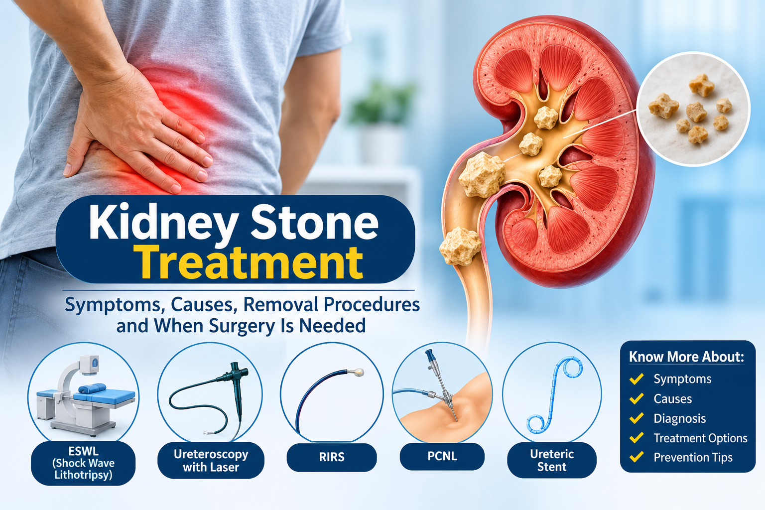
Kidney Stone Treatment: Symptoms, Causes, Removal Procedures and When Surgery Is Needed
A sudden cramping pain in your back or side that rises and falls in waves is hard to ignore. When it comes with blood in the urine, burning while passing urine or nausea, a kidney stone is a likely cause. The question most people ask next is simple: will I need an operation? Often, the answer is no. Kidney stone treatment ranges from fluids and pain control for small stones to laser procedures, shock wave lithotripsy and, for large or complicated stones, surgery. What decides the route is the stone’s size and position, whether it blocks the flow of urine, whether there is infection, and how well your kidney is working. This guide covers kidney stone symptoms, causes, tests, every major treatment option and the situations where surgery is the safer choice. If you are looking for kidney stone treatment at Zynova Shalby Hospital, the local care and booking details are near the end. Quick answer: Stones smaller than about 5 mm often pass on their own with fluids, pain relief and monitoring. Larger stones, stones that block the urinary tract and stones with infection usually need a procedure such as lithotripsy, ureteroscopy with laser, RIRS or PCNL. Severe pain with fever or chills is an emergency. What Are Kidney Stones? Kidney stones are hard deposits of minerals and salts that form inside the kidney when urine becomes too concentrated. Doctors also call the condition urolithiasis or nephrolithiasis. A stone can be as small as a grain of sand or large enough to fill part of the kidney. A stone that stays inside the kidney often causes no pain. Trouble starts when it moves into the ureter, the narrow tube that carries urine from the kidney to the bladder. The stone can block urine flow, and the kidney stretches as pressure builds behind it. That pressure is what causes the severe pain. Kidney Stone Symptoms Symptoms depend on the stone’s size and where it sits. The most common ones are: Sharp pain in the back, side, lower abdomen or groin that comes and goes in waves Pain that moves downward toward the groin as the stone travels Pink, red or brown urine (blood in the urine) Burning or pain while passing urine, a frequent or urgent need to go, or pelvic pressure, especially when the stone is close to the bladder Nausea and vomiting Cloudy or foul-smelling urine Fever and chills, which can signal infection Stone pain is often described as restless pain. People cannot find a comfortable position, unlike a pulled back muscle that eases when you shift or rest. Some stones cause no symptoms at all and are found on a scan done for another reason. Even then, they are worth discussing with a urologist, because a silent stone can start to move or grow later. Kidney Stone Causes and Risk Factors Stones form when urine carries more crystal-forming substances, such as calcium, oxalate and uric acid, than the available fluid can keep dissolved. Several things tip that balance: Drinking too little water, or losing a lot of fluid through sweat in hot, humid weather A diet high in salt, animal protein or sugar A personal or family history of kidney stones Obesity and diabetes Gout or high uric acid levels An overactive parathyroid gland (hyperparathyroidism) Repeated urinary tract infections Certain medicines, including some diuretics, anti-seizure drugs and antacids Inherited conditions such as cystinuria Types of Kidney Stones The type of stone shapes both treatment and prevention, which is why a stone that has passed should be saved for analysis. Stone type Usual background Useful to know Calcium oxalate and calcium phosphate Low fluid intake, high calcium or oxalate in urine Together these make up most stones Uric acid Acidic urine, gout, high-protein diet Some can be dissolved with medicine that makes urine less acidic Struvite Linked to urinary tract infections Can grow quickly into large, branched stones Cystine Inherited, rare Tends to come back, so long-term follow-up matters How Kidney Stones Are Diagnosed A urologist begins with your symptoms, medical history and a physical examination. Tests then confirm the stone and show whether it is causing a blockage or infection. Urine test: looks for blood, infection and crystals. Blood tests: check kidney function, calcium and uric acid. Ultrasound: uses no radiation and can show a stone in the kidney and swelling caused by blockage. It may miss small stones in the ureter. It is often preferred in pregnancy. Non-contrast CT scan: usually the most accurate test for a stone’s size and exact position. X-ray of the abdomen: sometimes used to track certain stones over time. Whether the stone is blocking urine flow often matters more for treatment than the stone’s size alone. How Stone Size and Location Affect Kidney Stone Treatment Size and position are the two facts that guide most decisions. The ranges below follow commonly used international urology guidance and are a general map, not a prescription. Your urologist may choose differently based on your scan and health. Situation What it usually means Ureter stone under about 5 mm, no infection, pain controlled Most pass on their own. Observation with fluids and pain relief is common. Ureter stone of 5 to 10 mm Roughly half pass. Doctors may observe for a limited time, offer medicine, and intervene if it does not move. Ureter stone over 10 mm Unlikely to pass. A procedure is usually needed. Kidney stone up to about 20 mm Shock wave lithotripsy or ureteroscopy with laser (including RIRS) are the usual options. Kidney stone over 20 mm, or a complex stone PCNL is generally the first choice. Any stone with infection or blockage Urgent drainage and treatment, whatever the size. Location matters too. Stones in the lower part of the ureter, near the bladder, pass more readily than stones higher up. In the kidney, larger stones in the lower pole respond less well to shock waves. Very dense stones also resist shock waves. Pregnancy, blood thinners, bleeding disorders, a single working

Breast Cancer Stages Explained: Stage 1 to Stage 4
Breast cancer stages describe how big a breast tumour is and how far the cancer has spread. Doctors number them from 0 to 4. A lower number generally means the cancer is smaller and more contained. Stage 4 means it has spread to distant parts of the body, such as the bones, lungs, liver or brain. If you or someone in your family has just received a diagnosis, the word ‘stage’ can feel heavy. It helps to know what it actually measures. The stage is a way for the care team to describe the cancer clearly and plan treatment that fits. It does not decide everything, and it is only one of several things your doctors look at. This guide explains breast cancer staging in plain language. It walks through stage 1 breast cancer, stage 2, stage 3 and stage 4, shows how doctors work out the stage, and describes how breast cancer treatment by stage is usually approached. Your own reports and your oncologist’s advice always come first. What does breast cancer staging mean? Staging is the process of finding out whether a cancer has spread and, if so, how far. The American Cancer Society (ACS) describes the stage as a measure of how much cancer is in the body, which helps show how serious it is and how best to treat it. You may see stages written in Roman numerals on your reports: 0, I, II, III and IV. They match the numbers 0 to 4 used here. As a rule, the lower the number, the less the cancer has spread. Letters add detail, and within a stage an earlier letter means a lower stage, so 2A sits below 2B. The stage is worked out from a physical exam, imaging tests and biopsies. Depending on the system used, breast cancer staging can bring together tumour size, lymph-node involvement, spread to distant organs, tumour grade and biomarker results (hormone receptor and HER2 status). How do doctors determine the stage of breast cancer? Most doctors use the TNM system from the American Joint Committee on Cancer (AJCC). Three letters carry most of the weight. T: tumour size T describes the main tumour and whether it has grown into the skin or the chest wall under the breast. Higher numbers mean a larger tumour or wider growth into nearby tissue. T1: 2 cm or less (about three quarters of an inch) T2: more than 2 cm and up to 5 cm T3: more than 5 cm T4: any size, growing into the chest wall or skin N: lymph nodes Lymph nodes are small, bean-shaped glands that filter fluid in the body. N tells you whether cancer has reached nearby nodes and how many are involved. N0: no cancer found in nearby lymph nodes N1: cancer in 1 to 3 lymph nodes under the arm, or very small spread to nodes near the breastbone found on sentinel node biopsy N2: cancer in 4 to 9 underarm nodes, or enlarged nodes near the breastbone N3: cancer in 10 or more underarm nodes, or in nodes below or above the collarbone M: distant spread M stands for metastasis, which means spread to distant organs. M0: no distant spread found M1: spread to distant organs such as the lungs, liver, bones or brain Clinical stage, pathologic stage and prognostic stage Your first stage is usually the clinical stage. It is based on the physical exam, imaging and biopsy. After surgery, the tissue that was removed is examined under a microscope, which gives the pathologic stage, also called the surgical stage. ACS notes that the cancer has sometimes spread further than the clinical stage suggested. So your stage can be refined after surgery or further tests. That is a normal part of the process. You may also hear the terms anatomic stage and prognostic stage. The anatomic stage uses only tumour size, lymph nodes and distant spread. The prognostic stage, part of the AJCC system used since 2018, combines those with grade, hormone receptor and HER2 results, and sometimes a gene test score. It also comes in clinical and pathologic versions. This is why your report can show a different stage from the one you would reach from size and nodes alone. Other findings that shape your stage and treatment plan Your pathology report will usually include these details: Grade describes how abnormal the cancer cells look and how quickly they are likely to grow. Low-grade cells look more like normal cells. High-grade cells look more abnormal and tend to grow faster. Grade and stage are different things, and people often mix them up. Hormone receptor status (ER and PR) shows whether the cancer cells carry receptors for the hormones estrogen and progesterone. When they do, hormone-blocking treatment can help. HER2 status shows whether the cells make too much of a protein called HER2. HER2-positive cancers can be treated with medicines that target that protein. Some people also have a gene test, such as the Oncotype DX recurrence score. In certain early-stage, hormone receptor-positive, HER2-negative invasive cancers, the result can help you and your doctor decide whether chemotherapy is likely to add benefit, and it can be taken into account in staging. It is not needed for everyone. Because these details are part of staging, two people with similar tumour sizes can end up with different overall stages. Ask your doctor about your own stage and what it means for you. Breast cancer stages at a glance, with usual treatment approaches The table gives a simplified description of each stage. It should not be used to determine an individual patient’s stage, which your doctors set from your reports. Stage Simplified description What it often means for planning Stage 0 Non-invasive cancer (such as DCIS). Abnormal cells have not broken out of the duct. Usually surgery, sometimes with radiation Stage 1 Small invasive tumour, about 2 cm

Knee Replacement Surgery: When Is It Needed and What Should You Know?
Most people don’t decide to look into knee replacement in one day. It builds slowly. First the knee feels stiff after sitting for a while. Then you start avoiding stairs, skipping long walks, or planning your day around your knee. At some point you begin to wonder whether surgery is the next step. There is no single sign that answers that question. Doctors look at how much your knee pain affects daily life, what your examination and scans show, and your overall health. This guide explains when knee replacement surgery is usually considered, how surgeons decide if it is right for you, and what you can try first. In short: knee replacement is usually considered when severe arthritis or joint damage keeps causing pain, stiffness or difficulty moving, and exercise, medicines, weight management or injections have not helped enough. Age alone does not decide it, and neither does any one symptom. What knee replacement surgery actually involves People often say the knee joint is “replaced”, but resurfaced is closer to the truth. The surgeon removes worn cartilage and a thin layer of bone from the ends of the thigh bone and shin bone, then fits metal components that recreate the joint surface. A medical-grade plastic spacer sits between them so the knee glides smoothly. The underside of the kneecap may also be resurfaced. In a total knee replacement, all the main surfaces of the joint are treated. In a partial knee replacement, only the damaged section is treated, which can suit some people whose arthritis is limited to one part of the knee and whose ligaments are healthy. Your surgeon decides which approach fits your knee after examining you and reviewing your scans. Why knees reach this point Most long-term knee pain comes from arthritis. Osteoarthritis is the most common type, a slow thinning of the cartilage that cushions the joint. It usually shows up after 50 but can appear earlier. Rheumatoid arthritis, where the immune system inflames the joint lining, and post-traumatic arthritis, which can follow a fracture or ligament injury, are the other major causes. As cartilage wears away, the bones rub against each other, and the result is pain, stiffness, swelling and sometimes a visible change in the shape of the leg. When knee replacement is usually considered Surgeons rarely look for one deciding symptom. They look at how much the knee limits your life and whether other treatment has stopped working. The American Academy of Orthopaedic Surgeons (AAOS) describes people who tend to benefit as those with: Severe pain or stiffness that limits everyday activities such as walking, climbing stairs and getting out of a chair, sometimes to the point of needing a cane or walker Moderate to severe knee pain at rest, including at night Long-lasting swelling and inflammation that do not settle with rest or medicines A knee that has bowed inward or outward Little improvement despite treatments such as anti-inflammatory medicines, injections or physiotherapy The UK’s NICE guideline on osteoarthritis frames it in similar terms. Referral for joint replacement is worth considering when knee symptoms substantially affect quality of life and non-surgical treatment has not worked or is not suitable. Notice what is missing from both descriptions. Neither depends on a particular age, and neither treats a scan as the whole story. Two people with similar X-rays can feel very differently, which is why the conversation always begins with how the knee affects your day. Symptoms worth getting checked You do not have to wait until the joint is badly damaged to see a specialist. An orthopaedic evaluation makes sense if: Knee pain has lasted several weeks and is not improving with rest and simple care Pain wakes you at night or bothers you even when you are sitting Swelling keeps returning The knee locks, catches or feels as though it might give way Stiffness is slowly increasing, or you can no longer fully straighten or bend the knee You have started avoiding stairs, longer walks or family outings because of your knee An evaluation is not a commitment to surgery. Often it ends with a plan built around exercise, weight management and medicines. If your pain flares mainly in the rainy months, our guide to monsoon joint pain and knee pain explains what is usually temporary stiffness and what deserves a closer look. Some symptoms should not wait. A knee that becomes hot, red and swollen, especially with fever, or sudden difficulty putting weight on the leg after a fall or twist, needs prompt medical attention. These can have causes other than arthritis. How surgeons decide whether you are a good candidate At a consultation, the surgeon takes a detailed history, checks how the knee moves, and assesses its stability, strength and the alignment of your leg. X-rays are reviewed, and sometimes blood tests or an MRI are added. Behind all of this, the surgeon is weighing a few practical questions: Do your symptoms and your scans tell the same story? Have you had a fair trial of non-surgical treatment? Is the damage confined to one part of the knee or spread throughout? Are other health conditions, such as diabetes, heart disease or kidney problems, well controlled? These can raise the risks of an elective operation, so they are usually addressed first. Are you ready for rehabilitation? Recovery takes regular exercise and some help at home for a few weeks. Age and weight deserve a straight answer. AAOS states that there are no absolute age or weight restrictions for total knee replacement. Most patients are between 50 and 80, but surgeons assess each person individually. NICE advises that age, smoking, other health conditions or obesity should not, on their own, rule someone out of referral. That said, a healthier weight and well-controlled conditions generally make surgery safer and recovery smoother. At Zynova Shalby Hospital, the orthopaedic team can work alongside the Diabetes 360 clinic and cardiology department when a patient’s wider health needs attention before surgery. What to try before surgery

Monsoon Joint Pain: Causes, Treatment, Prevention & When to See a Doctor
If your knees, shoulders or fingers seem to ache more once the rains set in, you are not imagining things. Joint pain during monsoon is one of the most common complaints in orthopaedic clinics between June and September. The same few lines come up every year: “It was fine all summer, then the rains started and my knee just stiffened up.” Or the classic: “Barish aayi toh dard badh gaya.” This article walks through why that happens, what actually causes monsoon joint pain, how to tell it apart from a genuine underlying joint problem, and what to do about it safely, at home and with medical help. Why Does Joint Pain Get Worse During Monsoon? Joint pain often feels worse during monsoon because of a combination of factors, not the rain by itself. Lower physical activity, prolonged indoor sitting, humidity, temperature drops and pre-existing joint conditions like arthritis can all add up to more stiffness and discomfort during the rainy season. Picture someone with mild knee arthritis who walks every evening through summer, takes the stairs, stays generally active. The rains begin, the evening walk gets skipped a few days in a row, the roads turn slippery, and without really deciding to, this person is now sitting far more than usual. That drop in movement alone is enough to make a joint feel stiffer, even before you factor in anything about the weather itself. It’s a pattern that shows up again and again in clinic, and it’s a large part of why joint pain in rainy season feels like a seasonal complaint, when really the underlying joint condition has usually been sitting there all along, just less active. Does Rain Actually Cause Joint Pain? Rain does not directly cause joint damage. Some people with existing joint conditions, particularly osteoarthritis or other forms of arthritis, do report more discomfort when the weather changes. But the exact reason is not fully understood, and it varies a lot from person to person. Weather is best thought of as a possible trigger, not a true cause. Researchers have looked at the weather and joint pain link for decades, and honestly, the results are mixed. Some people are clearly more sensitive to shifts in humidity or atmospheric pressure. Others notice nothing at all, even with a similar joint history. What most doctors agree on is simpler than either extreme: weather does not create new joint disease. It may just make an already sensitive joint more noticeable for a stretch of time. What About Atmospheric Pressure and Humidity? Atmospheric pressure and humidity come up a lot in this conversation, and they are worth a mention, carefully. The tissue around a joint may respond slightly to these shifts, particularly in people who already have some joint wear and tear. Treat this as a possible contributing factor, not a proven cause of joint damage. Rain, cold air or humidity does not wear down cartilage, and it does not create arthritis in a joint that was healthy to begin with. Common Causes of Joint Pain During the Rainy Season Here’s the thing worth remembering: causes of joint pain during monsoon are usually the same causes that exist all year round. Rainy weather can turn the volume up on symptoms, but it almost never acts alone. Common contributors include: Existing arthritis, including osteoarthritis, rheumatoid arthritis or other inflammatory joint conditions Osteoarthritis, the gradual wear of cartilage that cushions a joint, most common in the knees A previous joint injury, such as an old ligament tear or fracture, that becomes more sensitive with disuse Muscle weakness around a joint, particularly the muscles supporting the knee General joint stiffness from reduced movement Prolonged sitting during rainy days, at home or at work Increased body weight, which places extra load on weight-bearing joints like the knees and hips Tendon or ligament strain from sudden or unaccustomed activity Inflammatory joint conditions that flare up periodically regardless of season Sudden changes in activity level, such as going from very inactive to very active over a short period This is exactly why a doctor will ask several questions before agreeing that body pain during monsoon is purely weather-related. A proper history often turns up an underlying joint issue that was already there, just quieter, until the routine changed. Why Can Knee Arthritis Feel Worse During Monsoon? The knee is a weight-bearing joint that depends heavily on regular movement and strong surrounding muscles. When movement decreases, as often happens during monsoon, the knee can feel stiffer and less flexible. For someone who already has knee arthritis, this combination of reduced activity, muscle weakening and possible weather sensitivity can make arthritis pain during monsoon more apparent. Knee pain during monsoon shows up in a few recognisable patterns: Stiffness that is worse first thing in the morning or after sitting for a while Discomfort while climbing stairs or getting up from a low chair A feeling of heaviness or reduced flexibility in the knee Mild swelling around the joint after a long day of standing or walking Pain that improves somewhat once the joint is warmed up and moving For people without arthritis, occasional knee stiffness during monsoon is usually temporary and related to lower activity levels. For people who already have osteoarthritis knee pain, the same rainy season can bring a more noticeable increase in symptoms, sometimes described as an arthritis flare-up. Understanding Osteoarthritis in Simple Terms Osteoarthritis is a common joint condition where the smooth cartilage that cushions the ends of bones gradually wears down over time. Without this cushioning, movement can become stiffer and sometimes painful, particularly in weight-bearing joints like the knee. It develops gradually over years and is influenced by age, previous injury, body weight and joint use, not by a single season or one episode of rain. The Key Difference: Temporary Weather-Related Stiffness vs Persistent Joint Pain This distinction matters more than almost anything else in this article, so it is worth explaining clearly. Temporary weather-related stiffness tends to: Come on gradually

Heart Blockage Treatment: When Do You Need Medicines, Angioplasty or Bypass Surgery?
If a doctor has just told you or a family member about a heart blockage, your mind is probably racing with questions. Does this mean surgery right away? Will medicines be enough? Why did the doctor suggest angioplasty for one person and bypass surgery for another, even though both had blocked arteries? This confusion is common, and it is fair. Heart blockage treatment is not one-size-fits-all. Two patients with a similar looking blockage on a report can end up with completely different treatment plans, because treatment depends on much more than just a single number on an angiography report. This article explains, in simple language, how doctors actually think through this decision. It covers coronary artery disease, what a heart blockage means, how it is diagnosed, and how cardiologists choose between medicines, angioplasty with stent placement, and bypass surgery (CABG). What Does a Heart Blockage Actually Mean? The heart gets its own blood supply through coronary arteries. Over time, fatty deposits, cholesterol and other substances can build up inside these arteries. This buildup is called plaque, and the process is known as atherosclerosis. As plaque builds up, the artery becomes narrow. This narrowing is what people usually mean by a “heart blockage” or coronary artery blockage. When the artery is narrow enough to reduce blood flow to the heart muscle, it is called coronary artery disease (CAD). A blocked heart artery does not always cause noticeable symptoms right away. Some blockages are found only when a person undergoes tests for another reason, or during a routine health check. Symptoms That May Point to a Heart Blockage Coronary artery disease can show up differently in different people. Some common symptoms include: Chest pain, pressure, tightness or discomfort (often called angina) Breathlessness, especially during physical activity Unusual tiredness or fatigue that is not explained by exertion Sweating without an obvious reason Dizziness or lightheadedness Pain that spreads to the arm, shoulder, jaw, neck or back Not everyone experiences classic chest pain. Some patients, particularly women and people with diabetes, may only notice breathlessness or fatigue. A word of caution: if you or someone near you experiences sudden or severe chest pain, especially along with breathlessness, sweating, nausea, fainting or pain spreading to the arm or jaw, this can be a medical emergency. Do not wait to see if it passes and do not try to self-diagnose or self-treat at home. Seek urgent medical attention immediately. How Doctors Diagnose a Heart Blockage Before deciding on any treatment, a cardiologist needs a clear picture of what is happening inside the coronary arteries. Common tests include: ECG (Electrocardiogram): A quick, painless test that records the electrical activity of the heart. It can show signs of reduced blood flow or a past heart attack. Echocardiography: An ultrasound of the heart that shows how well the heart muscle and valves are working. Stress testing: Where appropriate, this test checks how the heart responds to physical exertion, which can reveal blockages that only cause problems during activity. CT coronary angiography: A non-invasive scan that gives detailed images of the coronary arteries and can help identify blockages without an invasive procedure. Coronary angiography: This is considered the most detailed way to look at the coronary arteries. A thin tube called a catheter is guided to the heart arteries, and a special dye is injected so that blockages show up clearly on X-ray images. Angiography helps the doctor understand exactly where the blockages are, how severe they are and how many arteries are affected. The results of these tests, taken together with your symptoms and overall health, guide the treatment decision. Why the Same Blockage Can Mean Different Treatments for Different People This is one of the most misunderstood parts of coronary artery disease treatment. Patients often ask why a 90 percent blockage in one person needs a stent, while a similar blockage in someone else is managed with medicines alone. The truth is that no single percentage of blockage automatically decides the treatment. Doctors look at several factors together, including: Location of the blockage: A blockage in a major artery, such as the left main coronary artery, is treated differently from a blockage in a smaller branch. Severity and how it affects blood flow: Two blockages that look similar on a scan can behave differently depending on how much they restrict blood flow. Number of arteries involved: Single artery blockage, two artery blockage and three artery or multi-vessel blockage often need different approaches. Symptoms: Whether you have chest pain during rest, during exertion, or no symptoms at all. Heart function: How well the heart muscle is pumping overall. Coronary anatomy: Whether the blocked segment is suitable for a stent, based on its shape, length and location. Other health conditions: Diabetes, kidney disease, prior heart attack, valve disease and other illnesses can influence the choice of treatment. Overall medical fitness: Age, general health and ability to tolerate a procedure or surgery. This is why heart blockage treatment must always be individualised. A blockage that needs bypass surgery in one patient may need only medicines and lifestyle changes in another, even if the percentage of narrowing looks similar on paper. Treatment Option 1: Medicines and Lifestyle Management Medicines are an important part of treatment for coronary artery disease. Depending on the severity and location of the blockage, symptoms, blood flow, heart function and overall health, your cardiologist may recommend medicines and lifestyle changes, either alone or alongside a procedure. Commonly used medicines for heart blockage may include: Antiplatelet medicines, to reduce the risk of clot formation Cholesterol-lowering medicines, to help slow further plaque buildup Blood pressure medicines, to reduce strain on the heart Medicines that reduce the heart’s workload and help relieve angina It is important to understand what medicines can and cannot do. Medicines for heart blockage help control symptoms, manage risk factors like high blood pressure, high cholesterol and diabetes, and reduce the risk of complications such as heart attack. However, medicines do not simply dissolve or remove

Dengue vs Malaria vs Leptospirosis: How to Tell Them Apart
If you’ve searched for lung cancer information before landing here, chances are you found a lot of generic content written for a Western audience. Pages that talk about smoking as the main cause, list symptoms in clinical language, and never once mention air pollution, chulha smoke, or gutka. That’s a problem, because lung cancer causes and symptoms in India don’t always match what’s written on international health sites. The causes are different, the age at diagnosis is different, and a large number of patients here have never touched a cigarette in their life. This guide covers the lung cancer risk factors relevant to Indian households, the lung cancer signs early stage patients tend to brush off as “just a cough,” how the lung cancer diagnosis process actually works here, and what lung cancer treatment in India looks like once you’re inside the system. This is educational information, not a diagnosis. If any of this sounds like what you or someone in your family is going through, please see a pulmonologist or oncologist rather than relying on an article to guide next steps. Why Lung Cancer Looks Different in India A few numbers worth knowing, drawn from lung cancer India statistics published by the Indian Council of Medical Research and the National Cancer Registry Programme: Lung cancer cases in India were projected to cross 81,000 in men and over 30,000 in women by 2025, a sharp rise from the last decade. Indian patients are typically diagnosed in their mid-50s, about ten years earlier than patients in Western countries. Roughly 75-80% of cases are caught only at stage 3 or 4, when the cancer has already spread beyond the lung. That’s the single biggest reason survival rates here remain lower than they should be. Lung cancer is no longer just a men’s disease in India. Rates among women are rising steadily in several major cities. None of this is meant to alarm you. It’s meant to explain why “wait and see” is the wrong approach with lung symptoms that don’t go away, and why catching this early makes such a dramatic difference to outcomes. Causes and Risk Factors: What’s Actually Driving This in India Smoking, But Not Only Cigarettes Tobacco remains the leading cause among all lung cancer risk factors, and India has close to 100 million adult smokers. But smoking here isn’t limited to cigarettes. Bidis, hookah, and smokeless tobacco (gutka, khaini, paan masala) all carry cancer risk, and smokeless tobacco in particular tends to get dismissed as “safer” than smoking. It isn’t, and it’s linked to oral and lung cancers both. Air Pollution This is where Indian risk factors diverge sharply from what global health sites talk about. Long-term exposure to PM2.5 pollution, the kind that spikes in Delhi-NCR, Mumbai, and most major Indian cities every winter, is a well-established driver behind air pollution lung cancer in India, including in people who have never smoked. If you live in a high-pollution city and have a persistent cough, that context matters. Biomass Fuel Exposure In many Indian households, especially in rural areas, cooking is still done using chulhas fired by wood, dung cakes, or crop residue. Years of exposure to this indoor smoke is a well-documented lung cancer risk, and it disproportionately affects women, since they’re usually the ones cooking. This single factor barely gets a mention on most lung cancer websites, despite being a real and significant cause here. Occupational Exposure Workers in construction, mining, and certain factory settings face exposure to asbestos, silica dust, and diesel exhaust, all linked to lung cancer risk. If your job involves years of exposure to dust or industrial fumes, it’s worth mentioning to your doctor even if you’ve never smoked. Genetic and Molecular Factors Roughly one in five lung cancer cases worldwide occurs in people who have never smoked, and the pattern of lung cancer in nonsmokers India appears to skew meaningfully higher among women here. A notable share of these cases are driven by specific gene mutations (EGFR being the most common in Indian patients), not tobacco at all. This matters clinically too, because these mutations respond to targeted drug therapies that wouldn’t necessarily be the first choice for a smoking-related cancer. Secondhand Smoke and Family History Living with a smoker for years raises risk even if you’ve never smoked yourself. A family history of lung cancer also modestly raises individual risk, though it’s a smaller factor than tobacco or pollution exposure. Early Signs That Are Easy to Ignore This is the part that costs the most lives, not because people don’t have symptoms, but because the early ones look so ordinary that nobody takes them seriously until much later. Symptoms worth paying attention to if they persist beyond 2-3 weeks: A cough that won’t go away, or one that changes character (gets worse, becomes dry when it used to bring up phlegm, or vice versa) Coughing up blood or blood-tinged mucus, even a small amount Breathlessness that seems out of proportion to your activity level Chest pain that gets worse with deep breathing, coughing, or laughing A hoarse voice that doesn’t clear up Unexplained weight loss or loss of appetite Recurring chest infections or pneumonia in the same area of the lung Persistent fatigue that isn’t explained by anything else going on in your life Individually, every one of these can be something far less serious. A lingering cough after a viral infection, or fatigue from a stressful few weeks at work, is common and usually not cancer. The pattern to watch for is persistence without an obvious cause. A cough that should have resolved in ten days and is still there after four weeks deserves a doctor’s visit, not another round of home remedies. When to See a Specialist As a general rule, see a pulmonologist if: A respiratory symptom (cough, breathlessness, chest pain) has lasted more than 3 weeks without improvement You’ve coughed up any blood, even once You’re a current or former smoker over 40 with any

Lung Cancer in India: Causes, Early Signs, Diagnosis and Treatment Options Explained
If you’ve searched for lung cancer information before landing here, chances are you found a lot of generic content written for a Western audience. Pages that talk about smoking as the main cause, list symptoms in clinical language, and never once mention air pollution, chulha smoke, or gutka. That’s a problem, because lung cancer causes and symptoms in India don’t always match what’s written on international health sites. The causes are different, the age at diagnosis is different, and a large number of patients here have never touched a cigarette in their life. This guide covers the lung cancer risk factors relevant to Indian households, the lung cancer signs early stage patients tend to brush off as “just a cough,” how the lung cancer diagnosis process actually works here, and what lung cancer treatment in India looks like once you’re inside the system. This is educational information, not a diagnosis. If any of this sounds like what you or someone in your family is going through, please see a pulmonologist or oncologist rather than relying on an article to guide next steps. Why Lung Cancer Looks Different in India A few numbers worth knowing, drawn from lung cancer India statistics published by the Indian Council of Medical Research and the National Cancer Registry Programme: Lung cancer cases in India were projected to cross 81,000 in men and over 30,000 in women by 2025, a sharp rise from the last decade. Indian patients are typically diagnosed in their mid-50s, about ten years earlier than patients in Western countries. Roughly 75-80% of cases are caught only at stage 3 or 4, when the cancer has already spread beyond the lung. That’s the single biggest reason survival rates here remain lower than they should be. Lung cancer is no longer just a men’s disease in India. Rates among women are rising steadily in several major cities. None of this is meant to alarm you. It’s meant to explain why “wait and see” is the wrong approach with lung symptoms that don’t go away, and why catching this early makes such a dramatic difference to outcomes. Causes and Risk Factors: What’s Actually Driving This in India Smoking, But Not Only Cigarettes Tobacco remains the leading cause among all lung cancer risk factors, and India has close to 100 million adult smokers. But smoking here isn’t limited to cigarettes. Bidis, hookah, and smokeless tobacco (gutka, khaini, paan masala) all carry cancer risk, and smokeless tobacco in particular tends to get dismissed as “safer” than smoking. It isn’t, and it’s linked to oral and lung cancers both. Air Pollution This is where Indian risk factors diverge sharply from what global health sites talk about. Long-term exposure to PM2.5 pollution, the kind that spikes in Delhi-NCR, Mumbai, and most major Indian cities every winter, is a well-established driver behind air pollution lung cancer in India, including in people who have never smoked. If you live in a high-pollution city and have a persistent cough, that context matters. Biomass Fuel Exposure In many Indian households, especially in rural areas, cooking is still done using chulhas fired by wood, dung cakes, or crop residue. Years of exposure to this indoor smoke is a well-documented lung cancer risk, and it disproportionately affects women, since they’re usually the ones cooking. This single factor barely gets a mention on most lung cancer websites, despite being a real and significant cause here. Occupational Exposure Workers in construction, mining, and certain factory settings face exposure to asbestos, silica dust, and diesel exhaust, all linked to lung cancer risk. If your job involves years of exposure to dust or industrial fumes, it’s worth mentioning to your doctor even if you’ve never smoked. Genetic and Molecular Factors Roughly one in five lung cancer cases worldwide occurs in people who have never smoked, and the pattern of lung cancer in nonsmokers India appears to skew meaningfully higher among women here. A notable share of these cases are driven by specific gene mutations (EGFR being the most common in Indian patients), not tobacco at all. This matters clinically too, because these mutations respond to targeted drug therapies that wouldn’t necessarily be the first choice for a smoking-related cancer. Secondhand Smoke and Family History Living with a smoker for years raises risk even if you’ve never smoked yourself. A family history of lung cancer also modestly raises individual risk, though it’s a smaller factor than tobacco or pollution exposure. Early Signs That Are Easy to Ignore This is the part that costs the most lives, not because people don’t have symptoms, but because the early ones look so ordinary that nobody takes them seriously until much later. Symptoms worth paying attention to if they persist beyond 2-3 weeks: A cough that won’t go away, or one that changes character (gets worse, becomes dry when it used to bring up phlegm, or vice versa) Coughing up blood or blood-tinged mucus, even a small amount Breathlessness that seems out of proportion to your activity level Chest pain that gets worse with deep breathing, coughing, or laughing A hoarse voice that doesn’t clear up Unexplained weight loss or loss of appetite Recurring chest infections or pneumonia in the same area of the lung Persistent fatigue that isn’t explained by anything else going on in your life Individually, every one of these can be something far less serious. A lingering cough after a viral infection, or fatigue from a stressful few weeks at work, is common and usually not cancer. The pattern to watch for is persistence without an obvious cause. A cough that should have resolved in ten days and is still there after four weeks deserves a doctor’s visit, not another round of home remedies. When to See a Specialist As a general rule, see a pulmonologist if: A respiratory symptom (cough, breathlessness, chest pain) has lasted more than 3 weeks without improvement You’ve coughed up any blood, even once You’re a current or former smoker over 40 with any

Best Orthopedic Hospital in Mumbai: 7 Things to Check Before Choosing One
If you or a family member has been told you need knee surgery, a hip replacement, spine treatment, or care for a fracture, the first real decision isn’t which procedure to choose. It’s which hospital to trust with it. A quick search for the best orthopedic hospital in Mumbai throws up dozens of names, star ratings, and claims of “world-class care,” and most of them sound the same on the surface. This guide is written for that exact stage of the decision, before you’ve picked a procedure or a surgeon, when you’re still figuring out how to choose an orthopedic hospital that won’t let you down. We’ll walk through the specific things worth checking, how Mumbai’s orthopedic care landscape looks right now, and where Zynova Shalby Hospital fits into that picture. Why the Hospital Choice Matters More Than People Realize Orthopedic outcomes depend on more than a skilled pair of hands in the operating room. Infection control protocols, the physiotherapy team that sees you the day after surgery, how quickly a nurse responds at 2 a.m., and whether the hospital has handled a case like yours before, all of that adds up to the result you actually get. all of these factors contribute to your overall outcome. Two patients with the same diagnosis, operated on with the same implant, Two patients with the same diagnosis who undergo surgery using the same implant can have very different recoveries depending on the hospital around them. That’s why searches like orthopedic hospital near me or trusted orthopedic hospital near me are only a starting point. Location gets you in the door. What happens after that is where the real comparison begins. What to Actually Check Before You Choose 1. Surgeon Experience and Specialization Ask how many procedures similar to yours the surgeon performs in a year. That number tells you more than how many years they’ve practiced. A best orthopedic doctor in Mumbai for knee replacements may not be the right fit for a complex spine case, and that’s worth checking before you commit. At a properly staffed multispeciality orthopedic hospital in Mumbai, you should find surgeons with distinct sub-specializations, joint replacement, trauma, spine, sports injury, instead of one generalist handling everything. 2. Accreditation and Infection Control NABH accreditation isn’t just a certificate on a wall. It means the hospital follows documented protocols for sterilization, operation theatre hygiene, and post-surgical infection monitoring. Before you commit, ask directly about the hospital’s surgical site infection rate. A hospital confident in its numbers will share them without hesitation. 3. Technology and Equipment Robotic-assisted and computer-navigated joint replacement systems help with implant precision, but they’re only useful when paired with a surgical team trained to use them well. When you’re checking a best joint replacement hospital in India, look at whether the technology is a genuine part of daily practice or a single showcase machine mentioned only in marketing material. 4. Post-Operative Care and Physiotherapy Recovery from a fracture, hip surgery, or spine procedure happens mostly after the operation is over. A hospital that starts physiotherapy within hours of surgery, and has dedicated rehabilitation staff rather than a shared general ward team, tends to produce faster, more reliable recoveries. This matters just as much for a best hospital for fracture treatment in Mumbai as it does for planned joint replacements. 5. Patient Outcomes and Reviews Orthopedic hospital ratings in Mumbai are easy to find online, but read past the star average. Look for specifics: how patients describe pain management, how quickly they were mobilized, and whether follow-up care matched what was promised before surgery. 6. Transparent Costing A trustworthy hospital gives you a clear cost breakdown, implant charges, room category, physiotherapy sessions, before the procedure, not after. If a hospital hesitates when you ask for numbers in writing, treat that as a warning sign rather than a formality you can skip. 7. Emergency and Trauma Readiness Fractures and accidents don’t wait for appointment slots. A hospital with round-the-clock trauma care and an orthopedic surgeon on call is a meaningful safety net, especially if you’re comparing options for a family member rather than a planned surgery. How Mumbai’s Orthopedic Hospital Landscape Looks Today Mumbai has no shortage of established names in orthopedic care, with large multispecialty and quaternary care centers spread across the city, each with their own strengths in joint replacement, spine surgery, or sports medicine. Most of these hospitals compete on similar ground: recognized accreditation standards, robotic-assisted surgery options, and senior consultants with international fellowships. What actually separates one hospital from another isn’t the technology. Most major centers now offer some form of robotic or computer-assisted surgery, so that alone stopped being a differentiator a while back. It comes down to how well the surgical team, nursing staff, and physiotherapy unit actually work together on your recovery, and whether you can get to follow-up appointments without it eating your whole day. Where Zynova Shalby Hospital Fits Zynova Shalby Hospital in Ghatkopar West is a NABH-accredited, 100-bed multispeciality hospital that has been part of Mumbai’s healthcare landscape since 2007, with a long-standing focus on high-volume joint replacement surgery. It has also positioned itself as a robotic surgery hospital in Mumbai, offering computer and robotic-assisted joint replacement as a routine part of its orthopedic program rather than a one-off feature. On patient review platforms, the hospital currently holds close to a 4.8 out of 5 rating from patient feedback, which is worth checking yourself before you rely on any single review site. For patients specifically searching for an orthopedic hospital in Ghatkopar or an orthopedic surgeon in Ghatkopar, here’s what the hospital brings to the checklist above: A dedicated orthopedic and joint replacement department, with surgeons handling primary and revision joint replacements, arthroscopic procedures, spine conditions, and fracture management, rather than treating orthopedics as a side service. Surgeons with focused sub-specializations, including robotic and computer-assisted joint replacement, so patients aren’t routed to a general surgeon for a specialized procedure. NABH accreditation, covering documented infection control and patient
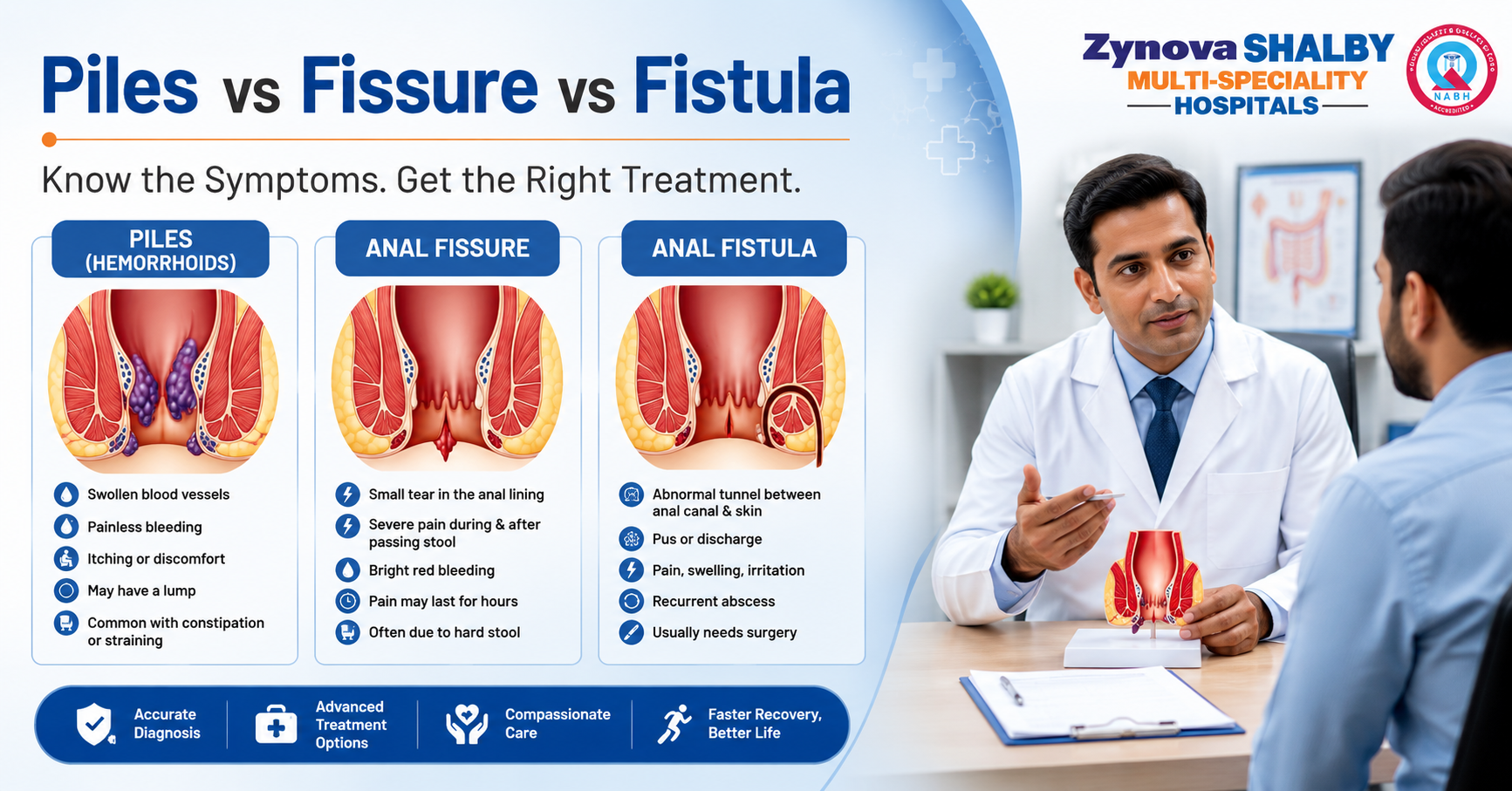
Anal Pain or Bleeding? It could be piles, a fissure, a fistula, or hemorrhoids.
Most people who notice blood in the toilet bowl, pain while passing stools, or itching near the anus assume it’s piles and either ignore it or reach for an over the counter cream. Sometimes that guess is right. Often it isn’t. Piles symptoms, anal fissure symptoms, and anal fistula symptoms overlap enough to fool almost anyone, and mixing them up is one of the most common reasons people stay in pain for months longer than they need to. If you’ve been trying to work out piles vs fissure vs fistula on your own, or searching things like anal pain causes, rectal bleeding causes, or blood in stool causes at 1 a.m. because you didn’t want to say any of this out loud to a doctor, this is written for you. We’ll walk through what actually separates these three conditions, what counts as a genuine emergency, where piles treatment without surgery and painless piles treatment actually apply, and where laser surgery becomes the realistic option. Towards the end, we’ll talk about what to look for before choosing a piles specialist in Mumbai, since a lot of clinics advertise the same laser packages without the same depth of experience in harder cases. There’s also an embarrassment factor nobody talks about enough. This part of the body isn’t something people bring up over dinner, sometimes not even with their own family doctor. That silence is exactly why fissures and fistulas get quietly misdiagnosed as piles for years. I’ve seen the pattern often enough while researching this that it’s worth saying plainly: if you’ve been putting this off, you’re not unusual, and it’s genuinely not worth waiting any longer. The reassuring part is that general surgery in Mumbai has grown enough in this space that proper anorectal care is now easy to find close to home, without having to explain yourself twice or sit through an uncomfortable waiting room conversation. Piles, Fissure, and Fistula Are Not the Same Thing Piles, also called hemorrhoids, are swollen blood vessels inside or around the anal canal. Everyone has these vascular cushions naturally. They only become “piles” when they enlarge, bleed, or push out during bowel movements. Straining, chronic constipation, prolonged sitting, and pregnancy are the usual hemorrhoids causes. Piles are graded 1 to 4 depending on how far they protrude, and that grading is really what decides whether you need a diet change, a banding procedure, or surgery. An anal fissure is a small tear in the lining of the anal canal, most often caused by passing a hard stool. It’s sharp, sometimes described as passing broken glass, and the pain can drag on for hours after you’ve actually finished using the toilet. Fissures under six weeks old are called acute. Ones that stick around longer become chronic, sometimes with a small skin tag forming near the tear (confusingly called a sentinel pile, which has nothing to do with actual piles). An anal fistula is a small tunnel that forms between the inside of the anal canal and the skin outside it, usually after an abscess, a pocket of infection, has drained on its own or been cut open. Unlike piles or fissures, a fistula almost never heals with medication or diet alone. There’s an actual physical tunnel that needs closing, whether through fistulotomy, a laser procedure, or techniques like LIFT or VAAFT depending on how complicated the tract is. Getting the right diagnosis changes everything about how it’s treated. A symptom checklist can point you in a direction, but it can’t replace an actual exam by someone who does this every day. Symptoms: What Points to What Symptom Piles Fissure Fistula Painless bleeding, bright red, on the stool surface Common Uncommon Rare Sharp pain during and after passing stool Rare, unless thrombosed Classic sign Sometimes Constant dull ache, worse when sitting Sometimes Sometimes Common Pus or foul smelling discharge No No Classic sign A lump or swelling you can feel externally Common (grade 3 to 4) Occasionally, a skin tag Common, an opening near the anus Anal itching causes on their own, no bleeding Common Occasionally Occasionally Recurrent boils or abscesses in the same spot No No Common If it’s just itching, with no bleeding or real pain, that’s usually piles, skin irritation, or a mild fungal issue rather than anything surgical. But persistent discharge, a lump that keeps returning, or an ache that’s there even when you’re not passing stool leans towards a fistula, and that needs an in person exam, not another week of home remedies. What Causes These Conditions in the First Place A quick word on prevention, since this comes up as often as treatment does. Piles and fissures share most of the same root causes: low fiber diets, not drinking enough water, sitting for long stretches at a desk, straining during bowel movements, and pregnancy related pressure on the pelvic floor. Fistulas usually start differently, from an anal gland infection that turns into an abscess, though poor hygiene, Crohn’s disease, and diabetes can all raise the risk of one forming or recurring. None of this means every case is preventable. Genetics play a role too, and plenty of people with a fibre rich diet and an active lifestyle still develop piles or a fissure. But if constipation is a recurring theme in your life, addressing that alone resolves a surprising number of mild cases before they ever need a procedure. When It’s Not Just Piles: Warning Signs You Shouldn’t Sit On A lot of anal pain causes are benign and settle with fiber, water, and a stool softener. But get seen urgently if you notice: Rectal bleeding that’s heavy, dark, or mixed into the stool rather than sitting on top of it Fever along with anal pain or swelling, which can point to an abscess that needs draining before it spreads A new lump that’s growing, especially with drainage or a bad smell Unexplained weight loss along with a change in bowel habits Bleeding in anyone over 45, or with